Visium HD and Visium spatial transcriptomics analysis
Whole-transcriptome spatial data at 2 µm resolution needs more than Space Ranger defaults. We turn Visium HD bins into cells, tissue domains and niches you can defend in review.
In short
OmicsQuest analyzes 10x Genomics Visium HD (2/8/16 µm bins) and standard Visium data from Space Ranger outputs or raw FASTQ. We run bin-level QC and tissue masking, segmentation-based aggregation of 2 µm bins into cells using H&E or immunofluorescence images, cell-type deconvolution (RCTD, cell2location), spatial domain detection, niche and neighborhood analysis, spatially variable genes and ligand-receptor signaling in tissue context. Pricing starts at $79/hour, and a pathology-aware biologist reviews every project.
Publication-quality figures
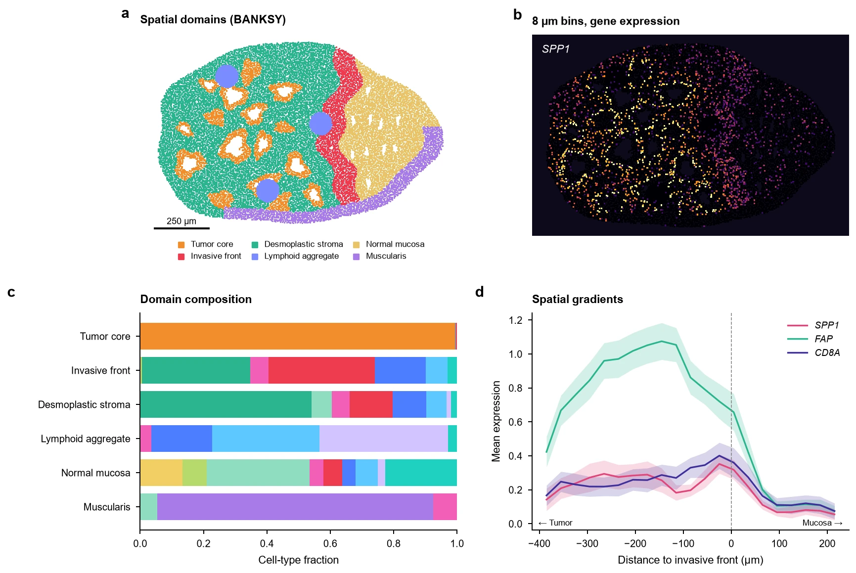
The kind of multi-panel figure we deliver, styled to journal specifications and revised until it is right.
Visium HD spatial analysis: analyses we run
Every analysis below is available from raw data or from your processed files. Pick the ones your question needs; we recommend a robust core set during scoping.
Data preparation and QC
- Space Ranger processing
- Reprocessing with correct probe set, image alignment and fiducial registration.
- Bin-level QC and tissue masking
- UMI and gene distributions at 2, 8 and 16 µm; removal of folds, edges and off-tissue bins.
- Diffusion and ambient signal assessment
- Quantify transcript spread beyond tissue boundaries before cell-level claims.
From bins to cells
- Nucleus and cell segmentation
- StarDist, Cellpose or 10x segmentation on H&E/IF, with expansion tuned to tissue type.
- Bin-to-cell aggregation
- Assign 2 µm bins to segmented cells (bin2cell-style), with quality filters per cell.
- Resolution choice
- Explicit comparison of 8 µm bins versus segmented cells for your biological question.
Cell identity in space
- Deconvolution
- RCTD, cell2location and Tangram, using matched or atlas scRNA-seq references.
- Label transfer to segmented cells
- Reference mapping with confidence scores and spatial sanity checks.
- Pathology-guided annotation
- Annotation checked against H&E morphology by a biologist.
Tissue architecture
- Spatial domains
- BANKSY, SpaGCN, GraphST or CellCharter domain detection.
- Spatially variable genes
- Moran's I, SpatialDE and nnSVG with appropriate multiple-testing correction.
- Neighborhood enrichment and niches
- Co-localization, neighborhood graphs and cellular niche composition (Squidpy).
- Tumor boundary and gradient analysis
- Distance-to-boundary expression gradients and invasive-front signatures.
Comparisons and signaling
- Multi-sample comparison
- Pseudobulk by domain or niche across samples and conditions, with replicate-aware statistics.
- Spatial cell-cell communication
- Ligand-receptor analysis restricted to spatial neighbors (COMMOT, LIANA+ spatial, NicheNet).
- Integration with Xenium or scRNA-seq
- Multi-platform integration on the same or serial sections.
What you receive
- QC report with tissue masks and bin metrics
- Segmented, annotated cell-level objects (SpatialData / AnnData / Seurat)
- High-resolution overlays on H&E and IF images
- Domain, niche and signaling figures sized for journals
- Code, software versions and methods text
Mistakes our experts avoid
- Treating 2 µm bins as cellsA 2 µm bin holds a fraction of a cell and a handful of UMIs. We aggregate bins to segmented cells or 8 µm bins depending on the question.
- Ignoring lateral diffusionTranscripts drift slightly during permeabilization, so very sharp boundaries can blur. We measure spread and avoid claims finer than the data supports.
- Deconvolving with the wrong referenceA reference missing a cell type forces its signal onto others. We check reference completeness and report unassigned signal.
- Comparing domains without replicatesOne section per condition is a case study, not a comparison. We design statistics around biological replicates.
- Clustering away tissue artifactsFolds, bubbles and edge effects form their own clusters. We mask them before analysis.
Tools and methods
We choose methods for your design, benchmark where it matters, and document versions for reproducibility.
- Space Ranger
- Loupe Browser
- SpatialData
- Squidpy
- bin2cell
- StarDist
- Cellpose
- RCTD
- cell2location
- Tangram
- BANKSY
- CellCharter
- COMMOT
- LIANA+
How a project runs
Scoping call
Thirty minutes, free. We learn the biology, the design and the deadline.
Capped estimate
A written plan with hours, deliverables and a cap. No surprise invoices.
Secure transfer and QC
Encrypted transfer, then a QC report and a go/no-go call within days.
Analysis with check-ins
Weekly calls with your duo team and interim figures you can steer.
Figures, methods, code
Publication-quality figures, methods text and reproducible code. Revisions are unlimited.
Projects we rescued
Frequently asked questions
What resolution should I analyze Visium HD data at?
It depends on the question. 8 µm bins are robust for domains and gene gradients. Segmented single cells (2 µm bins aggregated by nucleus segmentation) are better for cell-type neighborhoods and cell-level statistics. We usually deliver both and explain the trade-offs.
Do I need matched single-cell RNA-seq for Visium HD?
It helps, but it is not required. We can use high-quality public atlases as deconvolution references. Matched scRNA-seq gives the most accurate annotation for unusual tissues or disease states.
How much does Visium HD analysis cost?
Analysis is billed from $79/hour. A typical 4-8 section Visium HD project with segmentation, annotation, domains and niche analysis takes about 35-70 hours, and you get a capped estimate before we start.
Can you analyze FFPE Visium HD samples?
Yes. Most Visium HD projects we see are FFPE with CytAssist. We account for probe-based chemistry and RNA quality metrics in QC.
Further reading
Send us your Visium HD spatial data question.
Send a few lines about your experiment. A bioinformatician and a biologist will review it and reply with questions or a capped estimate. Analysis starts at $79/hour, and figure customization is unlimited.
- Free 30-minute scoping call
- Capped estimate before any billable work
- You own all data, code, figures and IP
Prefer email? contact@omicsquest.com